Similar
The Röntgen rays in medical work (1907) (14757181092)
Summary
Identifier: rntgenraysinmedi1907wals (find matches)
Title: The Röntgen rays in medical work
Year: 1907 (1900s)
Authors: Walsh, David
Subjects: X-rays Radiography X-Rays Radiography
Publisher: New York : William Wood
Contributing Library: Francis A. Countway Library of Medicine
Digitizing Sponsor: Open Knowledge Commons and Harvard Medical School
Text Appearing Before Image:
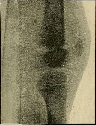
er epiphysis of the femur havebeen recorded, in which the diagnosis was confirmed by operation,autopsy or radiogram. He quotes Progressive Medicine, vol. iv.,.1900, p. 148. Separation of Lower Epiphysis of Femur. In an important article, Messrs. J. Hutchinson, jun., and H. L. Barnard describe four cases of this injury treated at the London Hospital, and mention sixteen others of which they have had personal knowledge. The injury is nearly always the result of forcible hyper- * Traumatic Separation of the Epiphyses, p. 97.f Practitioner, October, 1901, p. 395. MEDICAL AND SURGICAL APPLICATIONS 243 extension of the knee, and if displacement occurs, it is just as con-stantly a forward one of the epiphysis. By means of the Rontgenrays it was shown that in the extended position of the knee, evenwith an anaesthetic, reduction of the fragments was very difficult, ifnot impossible. The further important observation was made thatwith full flexion reduction was always easy. When treated in that
Text Appearing After Image:
Fig. 118.—Separation and Displacement of Lower Femoral Epiphysis.Hutchinson and Barnard. way the usual result was rapid recovery, with perfect movementof the knee, and without shortening or deformity of the leg (seeFigs. 118-120). Dislocation of the Epiphysis of a Metacarpal Bone. Mr. E. H. Herring, of Ballarat, found the above rare conditionin a boy of ten who had injured his hand by a fall from a tree. * Lancet, April 21, 1900, p. 1131. 16—2 244 THE RONTGEN RA YS IN MEDICAL WORK On examination there was found on the palmar surface of the righthand over the neck of the first metacarpal bone a small, immovable,rounded, hard nodule, of about the size of a pea. Several medicalmen diagnosed the case as one of exostosis. Treatment failed tokeep the nodule in place, and finally it was excised.
Tags
Date
Source
Copyright info


