Similar
Röntgen rays and electro-therapeutics - with chapters on radium and phototherapy (1910) (14758226345)
Zusammenfassung
Identifier: rntgenrayselectr00kass (find matches)
Title: Röntgen rays and electro-therapeutics : with chapters on radium and phototherapy
Year: 1910 (1910s)
Authors: Kassabian, Mihran Krikor, 1870-1910
Subjects: Electrotherapeutics X-rays Phototherapy Radiology Radiotherapy
Publisher: Philadelphia & London : J.B. Lippincott Company
Contributing Library: Francis A. Countway Library of Medicine
Digitizing Sponsor: Open Knowledge Commons and Harvard Medical School
Text Appearing Before Image:
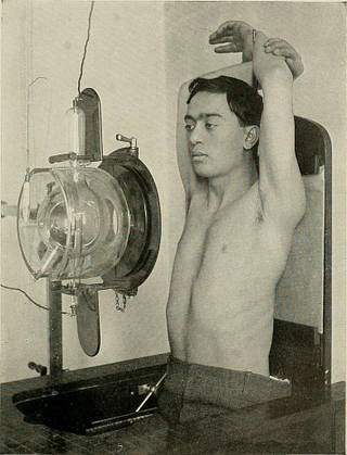
Reiniger,Gebberh SiSchal!, Enlangen.Fig. 182.—Levj-Dorns orthodiagraph for use in the recumbent posture. stylus and the tube. ISTow if a I is the apparent length of a foreign body,r I its real length, D the distance of the anticathode of the tube from theluminous screen, and d the distance of the object from the anticathode, cl r 1 X D the formula ^ will give the true distance of the foreign body from the luminous screen. The Levy-Dorn orthodiagraph is shown in Figs. 181 and 182. Theadvantage of this instrument lies in the fact that during the examina-tion of the heart the operator measures the vertical and horizontal axeson the scales. B. Skiagraphic Examination of the Heaet. The heart can be skiagraphed with the same technic as is applicableto the lung, but the former requires more precision in the position of thepatient, tube, distance, etc. The patient may be seated on a chair andthe plate placed either over the chest (sternum), in the anterior or
Text Appearing After Image:
Fig. 182A.—Lungs axd Heart f erect dorsal position.) This position may be more comfortable tosome patients. Tbe ventral view may be obtained by reversing the patients position. In taking stereo-scopic Eontgenograms of the chest, it will only be necessary to place the plate-changing box into thegrooves of the leaflet, and tube holder will be displaced by pulling out the chain, thus changing theposition of the anode 2% inches or 6 cm.
Tags
Datum
Quelle
Copyright-info


