Similar
Röntgen rays and electro-therapeutics - with chapters on radium and phototherapy (1910) (14571769637)
Zusammenfassung
Identifier: rntgenrayselectr00kass (find matches)
Title: Röntgen rays and electro-therapeutics : with chapters on radium and phototherapy
Year: 1910 (1910s)
Authors: Kassabian, Mihran Krikor, 1870-1910
Subjects: Electrotherapeutics X-rays Phototherapy Radiology Radiotherapy
Publisher: Philadelphia & London : J.B. Lippincott Company
Contributing Library: Francis A. Countway Library of Medicine
Digitizing Sponsor: Open Knowledge Commons and Harvard Medical School
Text Appearing Before Image:
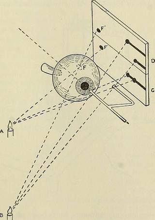
on, evenwhen carried out by making two exposures upon the same plate with thetube in different positions, or by making several separate exposures. Deutsche medicinische Wochenschrift, No. 18, 1897. 2 Medical Record, May 15, 1897. 3 Klin. Monatsblatt, Oct. 1897. * Wiener klin. Wochen., No. 7, 1898. 5 Trans. Amer. Ophth. Society, May, 1897. ® Diseases of the Eye, by Hansell and Sweet. THE CLINICAL APPLICATIONS. 285 The localizing apparatus designed by Sweet consists of two metalindicators, one pointing to the centre of the cornea and the other situatedto the outer cauthns at a known distance from the first. Two exposui-esare made in order to give different relations of the shadows of the indi-cators and of the body in the eyeball, one with the X-ray tube horizontalor nearly so with the plane of the indicators, and the other with the tubebelow this plane. The principle of the method may be understood from the perspec-tive drawing (Fig. 165). Eays coming from the light situated at A cast
Text Appearing After Image:
Fig. 165.—Principles of the method of localization. (Courtesy of Dr. Wm. M. Sweet.) shadows of two ball-pointed rods and an object in the eyeball, and givethe view shown on the surface C. In this instance the tube is in front ofthe vertical plane of the two indicators, and consequently the shadow ofthe centre ball wdll be thrown back of that of the outer ball. When thelight is carried below the plane of the two indicators, the shadows of thetwo rods are formed on the surface D, and the shadow of the foreignbody in the eye assumes a new position. If the distance of one of theindicating rods from the centre of the cornea is known, and the distance 286 ELECTEO-THEEAPEUTICS. between the two indicators is measured, the position of the metal in theeye may be determined, since the shadow of the foreign body preservesat all times a fixed relation to the shadows of the indicating balls, inwhatever position the light is placed. ^ Accurate localization requires that the axis of the eyeball sha
Tags
Datum
Quelle
Copyright-info


