Similar
American quarterly of roentgenology (1906) (14777160173)
Zusammenfassung
Identifier: americanquarterl01amer (find matches)
Title: American quarterly of roentgenology
Year: 1906 (1900s)
Authors: American Roentgen Ray Society
Subjects: Nuclear Medicine Radiography Radiology Radiology
Publisher: Pittsburgh : American Roentgen Ray Society
Contributing Library: Francis A. Countway Library of Medicine
Digitizing Sponsor: Open Knowledge Commons and the National Endowment for the Humanities
Text Appearing Before Image:
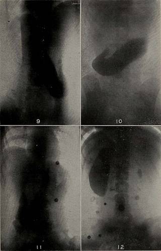
illedwith bismuth. Standing, the caudal pole came down tofour finger breadths below the navel. (No. 11.) Patient a girl, Aet. 14, lying on the belly. Dorso-ventral exposure. Notice the unusual shape of this stom-ach, with its screw-like twist and seeming break throughits middle from peristalsis. Its caudal pole is on a levelwith lower border of the 5th lumbar vertebra. Stand-ing, it comes down (orthodiagraphic examination) tofour finger breadths below the navel. Same patient on her back. Ventro-dorsal exposure.The stomach is somewhat higher than in No. 1. Thoughonly a few minutes have passed since she took the meal,bismuth shows in the small intestines. (No. 12.) Same patient on her right side. Dorso-ventral ex-posure. Notice that again this position seems to giveus the entire contour of the greater and lesser curva-tures, the pyloric portion, sphincter of the antrum, andlocation of pylorus. The right diaphragm is higher thanthe left, and the heart is displaced toward the right. (13.)
Text Appearing After Image:
To Illustrate the Presidents Address OF ROENTGENOLOGY. 11 Patient, a girl, Aet. about 28, on her belly. Dorso-ventral exposure. General direction of the stomach isdownward and to the left. Caudal pole is on a level withthe 5th lumbar vertebra. (No. 14.) Same patient on her back. Ventro-dorsal exposure.Fundus contains more mixture than with patient on herbelly, and caudal pole is still more to the left. (No. 15.) Same patient on her left side. Dorso-ventral expos-ure. Shape of stomach in this position is most extraordi-nary and resembles an almost straight piece of sausageof small cross section. The left diaphragm is higherthan the right. Heart is displaced toward the left. (16.) Same patient on her right side. Dorso-ventral ex-posure. The right diaphragm is now higher than the left.Heart displaced toward the right. Caudal pole very lowfor this position. Entire lower curvature covered withbismuth. Supernatant fluid sharply defined above thehorizontal line. Gas above the level of the fl
Tags
Datum
Quelle
Copyright-info


