Similar
The cyclopædia of anatomy and physiology (1847) (20199199504)
Summary
Title: The cyclopædia of anatomy and physiology
Identifier: cyclopdiaofana03todd (find matches)
Year: 1847 (1840s)
Authors: Todd, Robert Bentley, 1809-1860
Subjects: Anatomy; Physiology; Zoology
Publisher: London, Sherwood, Gilbert, and Piper
Contributing Library: MBLWHOI Library
Digitizing Sponsor: MBLWHOI Library
Text Appearing Before Image:
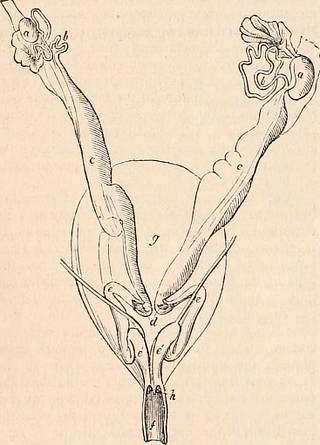
316 MARSUPIALIA. others partially divided ; but the divided por- tion in the latter is always that which is nearest the urethro-sexual passage. The true uterus is completely divided in all the Marsupial genera, and each division is of a simple elongated form, as in the Rodentia. The superadded complications in the female generative organs of the Marsupials, as com- pared with other mammals, are not then rightly attributable to the uterus, but to the vagina; and they are of such a nature as to adapt the latter to detain the foetus, after it has been ex- pelled from the uterus, for a longer period than in other Mammalia. These complications vary considerably in the different marsupial genera. On a comparison of the female organs in Didelphys dorsigera, Petaurus pygnueus, and Petaurus taguanoides, in Dasyurus viverrinus, in Didelphys Virgi- niana, in Macropus major, and Hypsiprymnus murinus, I find that the relative capacity which the uteri bear to the vaginae diminishes in the order in which the above-named species follow, while the size of the external pouch increases in the same ratio. In Didelphys dorsigera the uteri (Jig. 139, c, c,) rather exceed the unfolded vaginae in Fig. 139.
Text Appearing After Image:
Didelphys dorsigera. length. In most Marsupials the vaginae at first descend as if to communicate directly with the urethro-sexual passage; but in this small Opossum, in which the abdominal pouch consists of two slight longitudinal folds, and the young, as is implied by its trivial name, are transported by the mother on her back, each vaginal tube (e, e, ffig. 139,) after em- bracing the os tincae (d), is immediately con- tinued upwards and outwards, then bends downwards and inwards, and, after a second bend upwards, descends by the side of the opposite tube to terminate parallel with the extremity of the urethra (h) in the common or uro-genital passage (j'). In the Petauri, the vaginae, when unfolded, are a little longer than the uteri. On examining a specimen of the Pigmy Petaurist which had two very small young in the pouch, I found both the true uteri of three times the diameter of the same in an unimpregnated specimen ; but the vaginae were unaltered in size, indi- cating that the situation in which gestation takes place in this species is the same as in the Kangaroo. The vaginae, after receiving the uteri, descend close together half-way towards the commencement of the urethro-sexual pas- sage, but do not communicate together in this part of their course. From the upper part of these culs-de-sac they are continued upwards and outwards, forming a curve, like the han- dles of a vase, then descend, converge, and terminate close together, as in the preceding example. In Dasyurus viverrinus and Didelphys Vir- giniana, the mesial culs-de-sac of the vaginae descend to the urethro-sexual passage, and are connected to, but do not communicate with it. The septum dividing them from each other is complete, being composed of two layers which can be separated from each other, and which result indeed from the apposition and mutual cohesion of the vaginae at this part. In order to reach the common passage, each tube is con- tinued outwards from the upper end of the cul- de-sac, and forming the usual curve, terminates parallel to the orifice of the urethra. The vaginae in the Dasyures are smaller in propor- tion to the uteri than in the Virginian Opossum, but of a similar form. In another species, the Didelphys Opossum of Linnaeus, it would appear from the descrip- tion and figures of Daubenton,* that the septum of the mesial culs-de-sac of the vaginae was im- perfect ; but it is doubtful whether this inter- communication was not the result of parturition, or of an accidental rupture in the specimen ex- amined. If it should prove to be a specific difference of structure, it is an approximation to the condition of the female organs in the Phalangers, the Wombat, and the Kangaroos. In the Mucropus major the vaginas (jig. 138, e, e') preponderate in size greatly over the uteri (r, c') ; and, the septum (e") of the descending cul-de-sac being always more or less incom- plete, a single cavity (e) is thus formed, into which both uteri open ; but however imperfect the septum may be, it always intervenes and preserves its original relations to the uterine orifices (d, d). The foetus has been conjectured to pass into the urethro-sexual cavity by a direct aperture formed after impregnation at the lower blind end of the cul-de-sac, but I have not been able to discover any trace of such a foramen in two kangaroos which had borne young; and be- sides, I find that this part of the vagina is not continuous by means of its proper tissue * Buffon, Hist. Nat. torn. x. p. 279.
Tags
Date
Source
Copyright info


