Similar
The American journal of roentgenology, radium therapy and nuclear medicine (1906) (14754728244)
Summary
Identifier: americanjournroen06ameruoft (find matches)
Title: The American journal of roentgenology, radium therapy and nuclear medicine
Year: 1906 (1900s)
Authors: American Radium Society American Roentgen Ray Society
Subjects: Radiotherapy X-rays
Publisher: Springfield, Ill. C.C. Thomas
Contributing Library: Gerstein - University of Toronto
Digitizing Sponsor: University of Toronto
Text Appearing Before Image:
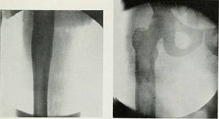
th .XniuKil Meeting of The American Roexigkn Ray Society, Saratoga Springs, N. Y., September 3-6. 1919. 594 Myxoma of Bone 595 to the middle of the shaft. The enlarge-ment was produced by the bulging out-ward of the cortical l^onc. There wasirregularity of outline of the cortexthroughout this area and thinning at theupper part just below the trochantermajor. The rest of the cortex in this areawas of normal thickness. No periostealbone formation was shown from the cortexor in the muscles. The plate made fivemonths after the first operation showed femur was made, and the patient wastaken to the hospital for operation severalweeks after being first seen. At this timethere was a tumor about the size of anorange over the anterior lateral surface ofthe upper third of the thigh. A long incisionwas made over the site of the tumor, whichwas found to be a bulging upward of thecrureus muscle from the shaft of the boneand cystic in character. On cutting intothe tumor it was found to contain gela-
Text Appearing After Image:
Kics. I AND 2. LiiiT Femur, Hip and Thigh one .Month befoke oilkaiion. practically the same conditions exceptthat there was thinning of the cortex withpointed bulging where the tumor haderoded the bone. The plate made im-mediately before amputation (sevenmonths after the first operation) showedmore bone destruction with periostealnew bone formation along the inner sur-face of the shaft extending into themuscles. Report of the Blood Exaniiiiatiou.—Wassermann negative; white cell count8,700 with 55 per cent polys., 33 per centlymphocytes, 3 per cent large monos., 2per cent eosinophiles, 6 per cent basophiles. A diagnosis of medullary tumor of the tinous material characteristic of a myxo-matous tumor. There was no blood in thecyst and grossly it did not look like sar-coma. From the gross examination bothDr. McCleary and I made a diagnosis ofmyxoma. The femur was exposed through-out the entire area of enlargement. A sinuswas found leading into the medullarycavity of the bone, and the co
Tags
Date
Source
Copyright info


