Similar
The American journal of roentgenology, radium therapy and nuclear medicine (1906) (14755568624)
Summary
Identifier: americanjournalo10ameruoft (find matches)
Title: The American journal of roentgenology, radium therapy and nuclear medicine
Year: 1906 (1900s)
Authors: American Radium Society American Roentgen Ray Society
Subjects: Radiotherapy X-rays
Publisher: Springfield, Ill. C.C. Thomas
Contributing Library: Internet Archive
Digitizing Sponsor: Internet Archive
Text Appearing Before Image:
, 1923) his visionis good and he is attending to his dailywork. It is an interesting speculationto what extent, if any, the disturbance ofhis pancreas followed the operation uponhis pituitary gland. Case 11. )\Iale, aged fifty-two. Referredby Dr. H. B. Stone, a nose andthroat specialist. Gives history of visualdisturbances for the past four years,not relieved bv glasses. Onset of visual Dr. Stone reported the following ocularfindings: Bitemporal hemianopsia. Cen-tral vision 20/30 20/50. Slight pallorof both nerve heads. Both well outlined.No choking. Terminal vessels tortuous. Patient was referred to Dr. GeorgeHeuer of Baltimore, who operated earlyin April, 1921, exposing the chiasmal regionby an intracranial approach. His reportfollows: We immediately exposed ^a\ery large hypophysial cyst which hadextended upward and directly compressedboth the optic chiasm and the mesialsurfaces of both optic nerves. We evac-uated the cyst, which contained a bloodygrumous material such as we usuallv
Text Appearing After Image:
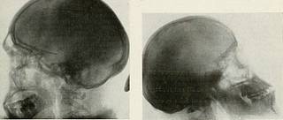
Fig. 2. Case II. Flour of sella extremely thin. Dorsumsella destroyed. Operative findings, cystic adenomaof pituitary. symptoms gradual. Headaches, bitemporalin character, worse at night, though forthe past several months have been better.Began putting on fat six years ago. Sometingling in extremities. First seen on August 14, 1920, at whichtime the sella was reported as being verydeep and large, the floor and the clinoidprocesses not easily made out. From theroentgen findings, a tumor of the hypoph-ysis was suggested at this time. The patient was told to return forfurther examination, but did not dothis until March, 1921, at which time thedorsum sella was completely destroyed.The floor of the sella was extremely thinand some encroachment on the sphenoidalsinus was noted. A roentgen diagnosis ofhypophysial tumor was made. Fig. 3. Case HI. Acromegaly. F\treiutl\ large sella.No destruction or thinning of floor. Large frontalsinus and inferior maxilla. find in degenerated adenomata of th
Tags
Date
Source
Copyright info


