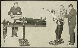
Röntgen rays and electro-therapeutics - with chapters on radium and phototherapy (1910) (14735220116)
Summary
Identifier: rntgenrayselectr00kass (find matches)
Title: Röntgen rays and electro-therapeutics : with chapters on radium and phototherapy
Year: 1910 (1910s)
Authors: Kassabian, Mihran Krikor, 1870-1910
Subjects: Electrotherapeutics X-rays Phototherapy Radiology Radiotherapy
Publisher: Philadelphia & London : J.B. Lippincott Company
Contributing Library: Francis A. Countway Library of Medicine
Digitizing Sponsor: Open Knowledge Commons and Harvard Medical School
Text Appearing Before Image:
xpiration it rests on the diaphragm, its long axis forming anacute angle with the imaginary median line of the thoracic cavity. Ininspiration the heart moves downward and toward the median line ; theright border of this organ is plainly seen to the right of the sternum, thelarger or left part of the heart is seen to the left of the sternum,—i.e.,the long axis of the heart forms with the median line during expirationa less acute angle than during an inspiratory effort. During inspirationthe transverse diameter of the heart is slightly decreased in length, atthe same time the number of pulsations are lessened. In expiration,after the diaphragm has discontinued tugging on the heart, the transversediameter is again increased, as is also the amplitude of its pulsations.The general contour of the organ can be more easily seen during inspira-tory periods than in the expiratory, because the lungs, being filled to theircapacity, are more transparent to the rays, thus offering a more striking
Text Appearing After Image:
APPLICATION OF THE X-EAYS. 321 contrast. The cardiac outline may be readily differentiated by means ofthe ingenious artifice of Dr. Disan.* By this method the outline of anormal heart is traced on the chest by fixing with adhesive strips a copperwire. A fluoroscopic examination is then made in the following way :At first the greatest strength of current obtainable from the apparatusis turned on. The observer looks through the fluoroscope and gets thechief landmarks of the chest, such as the scapula, ribs, spine, diaphragm,and upper convex border of the liver, the wire being at the same time infull view. The current is now reduced until the heart becomes more dis-tinctly visible. The fluoroscope is applied to a spot marked at the leftof the spine, corresponding to the fourth intercostal space in front of thechest. Any alterations in the shape of the heart can thus be easilydemonstrated. The shadows of the pulmonary vessel and in many instances the venacavce can be recognized if the che
Tags
Date
Source
Copyright info


