Similar
Practical points in the use of X-ray and high-frequency currents (1909) (14754437531)
Summary
Identifier: practicalpointsi00judd (find matches)
Title: Practical points in the use of X-ray and high-frequency currents
Year: 1909 (1900s)
Authors: Judd, Aspinwall
Subjects: X-rays Electrotherapeutics Radiography X-Rays Radiography
Publisher: New York : Rebman Company
Contributing Library: Francis A. Countway Library of Medicine
Digitizing Sponsor: Open Knowledge Commons and Harvard Medical School
Text Appearing Before Image:
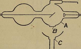
tube. When adjusted fora certain parallel spark gap, if its resistance becomes higher than theparallel spark gap it was set for, the spark will pass between Aand B so that the current is conducted into the small extensiontube having a chemical which, when the electricity passes through it,gives off a gas which lowers the resistance of the tube. When usingthe tube as a pointer, as shown on Fig. 37, it is necessary to turn thecurrent off and keep moving the pointer A a little closer or a littlefurther away according to the desired regulation of the tube. When,however, the tube is connected as shown by Fig. 46, then the operatordoes not have to shut off the current, as the spark gap is adjusted byraising or lowering the rod E of the middle spark gap. X-Ray Tubes 71 soda glass, which allows the X-ray to passthrough it easily. The rest of the bulb, being oflead glass, does not permit the X-ray to passthrough. There are lead glass shields madewhich fit on to the projection, so that the rays
Text Appearing After Image:
Fig. 38.—A represents a bulb of lead glass. B a window of sodaglass which allows X-rays to be passed through it. C a lead glassshield which fits on to the projection of the main tube, thus confiningthe rays to an area equal to the opening at the end of C. This tubewas designed by Dr. H. G. Piffard, and is intended to protect theoperator as well as the parts of the patient it is desired not to ray. are confined entirely to the area to be treated.This tube is made self-regulating. (See Fig.38.) A modification of this is known as the Cornelltube, designed by Dr. Albert Geyser. It is essen-tially a very small Piffard tube, but is used in adifferent way. The soda glass window is keptin absolute contact with the part to be treated.(See Fig. 39.) English Derma Tube. This is practicallythe original Crookes tube, having a handle at- 72 X-Ray and High-Frequency Currents tached to it and a shield to regulate the areatreated. This is also brought in contact withthe part to be treated. The X-ray
Tags
Date
Source
Copyright info


