Similar
Pathology and treatment of diseases of women (1912) (14758654596)
Summary
Identifier: pathologytreatme00mart (find matches)
Title: Pathology and treatment of diseases of women
Year: 1912 (1910s)
Authors: Martin, August Eduard, 1847- Jung, Ph. (Philipp Jacob), 1870-1918
Subjects: Gynecology Gynecology
Publisher: New York : Rebman company
Contributing Library: Francis A. Countway Library of Medicine
Digitizing Sponsor: Open Knowledge Commons and Harvard Medical School
Text Appearing Before Image:
. (Abdominal operation.) mata, enucleation is executed by opening the peritoneum and the capsuleat the apex of the tumor (Fig. 120). If several myomata have formed independently of each other, thenfrom the bed of the first enucleated tumor the second myoma is incised.If the tumors are too far apart from each other separate incisions aremade at such points on the surface as appear suitable. I seize thetumors with bullet- or Muzeux forceps; the introduction of drill in-struments only retards the process of the operation. The tumor, now, is PATHOLOGY OF THE VAGINA AND UTERUS 263 pulled upward and peeled out with the fingers. Stronger bands, connect-ing the growth with the capsule of the tumor are cut with knife or scis-sors. The bed of the tumor mostly does not bleed. The hemorrhagestops, at all events, if the assistant presses the cervix with both handsfrom the sides. Otherwise use artery clamps. The bed of the tumor issmoothed as much as possible, thereby the capsule is removed in most
Text Appearing After Image:
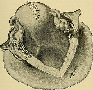
Fig. 122.—The uterus wound is sutured. cases. Henkel196 has recommended the systematic excision of the capsule.The uterine mucosa is examined thoroughly after opening of the uterinecavity. Abrasio mucosae through the existing opening, and eventuallyresection of the superfluous parts of the mucosa. Closure of the uterinecavity by a continued submucous catgut stitch (Fig. 121). Suturing ofthe bed of the myoma with continued sutures in rows. Coaptation ofthe wound margins by a continuous catgut stitch (Fig. 122). The en-tire body of the uterus is palpated for myoma-nuclei. Diseased ovariesand tubes are removed or resected. Examination - of the, peritoneal 264 DISEASES OF WOMEN cavity for changes previously unnoticed. Replacing- of the uterus,cleansing of the abdominal cavity, suturing of the peritoneum with con-tinued suture after placing a sterile cloth on the intestines which tendto press forward, which is removed, before the last stitch through theupper end of the peritoneal wound i
Tags
Date
Source
Copyright info


