Similar
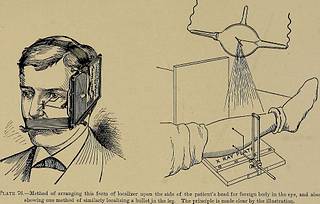
A system of instruction in X-ray methods and medical uses of light, hot-air, vibration and high-frequency currents - a pictorial system of teaching by clinical instruction plates with explanatory text (14570338528)
Summary
Identifier: systemofinstruct00mone (find matches)
Title: A system of instruction in X-ray methods and medical uses of light, hot-air, vibration and high-frequency currents : a pictorial system of teaching by clinical instruction plates with explanatory text : a series of photographic clinics in standard uses of scientific therapeutic apparatus for surgical and medical practitioners : prepared especially for the post-graduate home study of surgeons, general physicians, dentists, dermatologists and specialists in the treatment of chronic diseases, and sanitarium practice
Year: 1902 (1900s)
Authors: Monell, S. H. (Samuel Howard), d. 1918
Subjects: Vibration X-rays Diagnosis, Radioscopic Thermotherapy Electrotherapeutics X-Ray Therapy Vibration Diagnosis
Publisher: New York : E.R. Pelton
Contributing Library: Francis A. Countway Library of Medicine
Digitizing Sponsor: Open Knowledge Commons and Harvard Medical School
Text Appearing Before Image:
Plate 75.—Bullet Located by Mr. Banels Method. Three distinct indices were fixed tothe patient. The first was a small cross of tin-foil, which is readily visible in the negative,but can only be detected in the print by the white cross rising from the lines by which it wasmarked out. No use was made of this index. The third index, the only one used, was a nar-row strip of lead fixed between two prominent marks left by the stitches near the outer partof the gluteal fold. The patient lay on his back so as to about half cover a 10 x 12 plate,leaving plenty of space on which to stand the localizing cylinders. Two exposures on thesame plate were made, each three minutes, with electrolytic interrupter. The position ofthe bullet having been ascertained the patient was made to again lay on his back on a draw-ing-board. A mark was made on the skin at the indicated height, and the position of thebullet located. (Rebman, Ltd.)
Text Appearing After Image:
'
Tags
Date
Source
Copyright info


