Similar
A system of instruction in X-ray methods and medical uses of light, hot-air, vibration and high-frequency currents - a pictorial system of teaching by clinical instruction plates with explanatory text (14570272120)
Summary
Identifier: systemofinstruct00mone (find matches)
Title: A system of instruction in X-ray methods and medical uses of light, hot-air, vibration and high-frequency currents : a pictorial system of teaching by clinical instruction plates with explanatory text : a series of photographic clinics in standard uses of scientific therapeutic apparatus for surgical and medical practitioners : prepared especially for the post-graduate home study of surgeons, general physicians, dentists, dermatologists and specialists in the treatment of chronic diseases, and sanitarium practice
Year: 1902 (1900s)
Authors: Monell, S. H. (Samuel Howard), d. 1918
Subjects: Vibration X-rays Diagnosis, Radioscopic Thermotherapy Electrotherapeutics X-Ray Therapy Vibration Diagnosis
Publisher: New York : E.R. Pelton
Contributing Library: Francis A. Countway Library of Medicine
Digitizing Sponsor: Open Knowledge Commons and Harvard Medical School
Text Appearing Before Image:
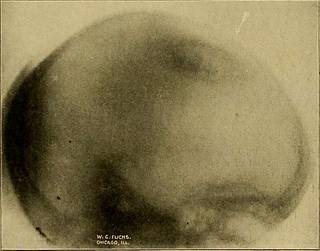
Plate 52 (1).—Cystic tumor in the brain in child who was blind and partially paralyzed.Arrow in upper left-hand corner points to the tumor. The plate below shows the lateralcross-section of same case, while this plate gives the antero-posterior view. Compare bothradiographs. The reduced half-tone loses much of the original negatives.
Text Appearing After Image:
Platk 52 (2).—Lateral view of above case of cystic tumor in the brain of child,operated on and recovered, verifying the radiographs. Case THE AUTHOES DISTOETION LANDMARK 209 X-ray work may be considered accurate when it attains its object.In some pictures tlie trained eye of tlie expert observer can note ata glance tbe slant of the shadows and deduce from them the positionof the tube without a registering landmark, but the adoption of astandard landmark to certify on the plate the relation of the axis ofthe rays to the plane of the part examiaed will do much to removedoubt as to how the anatomical relations of the recorded shadowsshould be interpreted. For this purpose, when needed, the authorhas devised a pair of thin steel circles mounted rigidly on a supportas shown in the Instruction Plates. As the majority of radiographsare made on films not smaller than 8 x 10 the base circle is seven inchesin diameter. This is large enough to easily embrace all the main diag-nostic field on a
Tags
Date
Source
Copyright info


