Similar
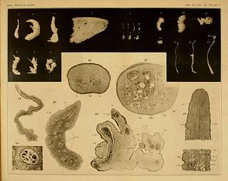
A Plate of 'Plerocerus prolifer' (= Sparganum proliferum) in Ijima (1905)
Summary
I. Ijima.
ON A NEW CESTODE LARVA PARASITIC IN MAN.
PLATE.
Explanation of figures.
Plerocercoides prolifer.
Fig. 1. A vertical slice of the skin and subdermal tissues taken from the left thigh of the patient, showing numerous encysted Plerocercoides proiifer in situ. Hardened in alcohol. Above, the epidermis. From some of the cysts the worm had fallen out. Natural size.
Fig. 2 a-g. Seven separate pieces of the worm taken from a single large cyst. Magnified li times. Photographed after fixing with corrosive sublimate, a-c, simple Plerocercoids. d, a strongly constricted piece of the worm (with involuted head ?). e-g, irregularly shaped pieces budding out heads.
Figs. 3-15. Worms in various shapes; all drawn from fixed specimens, magnified 4 times.
Fig. 3. A specimen of simple Plerocercoid shape, with the extreme head-end invaginated.
Fig. 4. Plerocercoid bearing a branch-like supernumerary head on one side.
Fig. 5. A similar specimen, bearing two supernumerary heads and strongly constricted in the middle.
Fig. 6. A specimen with a branch-like bud ; the terminal head, either not present or strongly withdrawn.
Figs. 7 and 8. Irregular-shaped specimens with numerous heads formed by budding.
Figs. 9 and 10. Contracted specimens, either without the head or with the same strongly wrthdrawn.
Fig. 11. A specimen, irregularly coiled and with tubercle-like protuberances.
Fig. 12. A piece constricted off from the hind parts of a Plerocercoid. Invaginated at both ends.
Fig. 13. A Plerocercoid greatly stretched out, but with the extreme head-end still retracted.
Fig. 14. A Plerocercoid with the terminal head either lost or strongly retracted, but with a greatly outstretched head-bud.
Fig. 15. A Plerocercoid moderately stretched out and with irregularities of contour in the anterior parts.
Fig. 16. Cross-section through the anterior part (head region) of a Plerocercoid. Magnified 100 times. %., lateral nerves, ex., excretory vessels in section. The black dots represent partly nuclei and partly longitudinal muscular fibers in section.
Fig. 17. Cross-section through the posterior part of a Plerocercoid. Magnified 100 times, cal., calcareous bodies, ex., excretory vessels. mus., bundles of longitudinal muscular fibers, which, in many other parts, are represented by the larger black dots. r. n., reserve nutritive-matter in capsule.
Fig. 18. Head-end of a Plerocercoid fully stretched out, showing the simply rounded tip. Drawn from a specimen clarified with glycerine, 30 times magnified. Excretory vessel in part strongly swollen on account of the stowing of the liquid contents.
Fig. 19. A Plerocercoid pressed under glass; over-stained with carmine and afterwards bleached with caustic potash. Black dots represent well-stained calcareous bodies, which are absent in the head region. Reserve nutritive-matter (r. n.) in the form of numerous balls. A pair of excretory vessels (ex.) in the anterior parts. Magnified 30 times.
Fig 20. A section through an irregular-shaped piece bearing a number of buds or heads, parts of which are seen in two places (A.). Other lettering as in fig. 17. Magnified 50 times.
Fig. 21. A horizontal section through the nearly fully evaginated head-end of a Plerocercoid. Lettering as in fig. 17. Magnified 50 times.
Fig. 22. A horizontal section through the hind parts of a Plerocercoid. Lettering as in fig. 17. Magnified 50 times.
Fig. 23. Section of a worm-cyst lying in the subdermal connective tissue. About 8 times magnified.
Date
Source
Copyright info


