Similar
A manual on foot care and shoe fitting for officers of the U.S. Navy and U.S. Marine corps (1920) (14597413648)
Summary
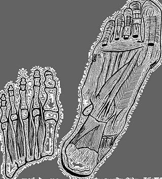
Left: Muscles controlling lateral toe action Right: Deep muscles of foot. Muscles and tendons controlling toe action
Identifier: cu31924031243045 (find matches)
Title: A manual on foot care and shoe fitting for officers of the U.S. Navy and U.S. Marine corps
Year: 1920 (1920s)
Authors: Mann, William Leake Folsom, Spencer Augustus, joint author
Subjects: United States. Navy United States. Marine Corps Foot Boots Footwear Shoes
Publisher: Philadelphia, P. Blakiston's Son & Co
Contributing Library: Cornell University Library
Digitizing Sponsor: MSN
View Book Page: Book Viewer
About This Book: Catalog Entry
View All Images: All Images From Book
Click here to view book online to see this illustration in context in a browseable online version of this book.
Text Appearing Before Image:
tfl lU I, « w d K -•-• V .^ 0) .rt +J + rf S H 0) g iH l2 ^ r S ^ S w g o § o B gt^-^ ^ „wC J, ■3 Sj -a ^ ^a ^«sS « 1-1 o o ^ « H - ■ • o m M g I—I -J 2 +* D u d 01 til ANATOMY AND PHYSIOLOGY II The longitudinal arch, on the inner side of the foot extendsfrom the heel bone (Os Calcis) to the distal end of the first
Text Appearing After Image:
Fig. 3.—Muscles con- Pig. 4.-e-Deep muscles of foot. Muscles trolling lateral toe action. and tendons controlling toe action. (C««-(Cunningham.) ningham.) metatarsal bone. (See illustration No. 2.) Thig is definitelyformed by the inherent structural concavity of the bones held 12 FOOT CARE AND SHOE FITTING among themselves by ligaments and supported from below bydeveloped muscle layers. The anterior arch is formed by the distal ends of the meta-tarsal bones. (SeeillustrationNo. s.) The muscular develop-ment concerned in sustaining this arch is not so great as in thelongitudinal. A B Fig. 3.—Cross section of feet showing metatarsal bones forming anterior arch. A shows formation of anterior arch by dit.tal ends of metatarsal bones.Note convexity of instep, dotted line indicating integrity of arch and con-cavity formed on the plane C. B shows fallen anterior arch. Note flat or convex instep, dotted line andabsence of concavity on the plane C. A tripod is formed by the structure of
Note About Images
Please note that these images are extracted from scanned page images that may have been digitally enhanced for readability - coloration and appearance of these illustrations may not perfectly resemble the original work.'
Tags
Date
Source
Copyright info


