Similar
The Dental cosmos (1907) (14781479395)
Zusammenfassung
Identifier: dentalcosmos4919whit (find matches)
Title: The Dental cosmos
Year: 1907 (1900s)
Authors: White, J. D McQuillen, J. H. (John Hugh), 1826-1879 Ziegler, George Jacob, 1821-1895 White, James William, 1826-1891 Kirk, Edward C. (Edward Cameron), 1856-1933 Anthony, L. Pierce (Lovick Pierce), b. 1877
Subjects: Dentistry Dentistry
Publisher: Philadelphia, S. S. White Dental Manufacturing Co
Contributing Library: Yale University, Cushing/Whitney Medical Library
Digitizing Sponsor: The College of Physicians of Philadelphia and the National Endowment for the Humanities
Text Appearing Before Image:
of themandible was extracted, and a number ofnecrotic pieces of bone were removedfrom the horizontal and descending rami,the cavity being thoroughly curetted andpacked with iodoform gauze. An area ofabout one and one-eighth inches of thelower end of the descending ramusand of the posterior part of the horizon-tal ramus and angle was thus removed.On account of the removal of this amountof bone, the jaw naturally deviated atonce to the left. To overcome this, aretaining appliance of rubber similar toa double-arch interdental splint was sub-sequently made, to hold the mandible incorrect articulation with the maxilla andto secure a close bite. The patient left the hospital June 24,1906. Since then he has worn the retain-ing appliance for three and a half orfour months about as diligently as thisclass of hospital cases may be expected todo; at that time he stopped wearing it,as he found that he had a good articula-tion and could use the mandible freely.On the following October the area re-
Text Appearing After Image:
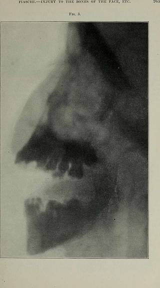
266 THE DEXTAL COSMOS. iDoved from the mandible was found tohave been completely regenerated, andwas firm and solid. The deviation of themedian line of the mandible from that ofthe maxilla amounts to about one-halfthe width of the lower central incisor,whereas immediately after the operationit was almost three-quarters of an inch.An X-ray plate (Fig. 3) taken at thistime shows the area of regenerated bonevery plainly, and also a faint line acrossthe descending ramus, where apparentlycalcification has not yet become com-plete. The interior of the mouth ishealthy with the exception that the boneat the angle does not on bimanual palpa-tion feel as thick as on the right side,there being in appearance no differencebetween the two sides. Bacteriologic ex- amination for the bacillus of Eberth wasnegative. I would call your attention to the ex-ceedingly fine result Dr. Hartley has ob-tained in operating on the case fromwithin the mouth, whereas in hospitals,cases of this class are generally t
Tags
Datum
Quelle
Copyright-info


