Similar
Röntgen ray diagnosis and therapy (1904) (14778055723)
Zusammenfassung
Identifier: rntgenraydiagn00beck (find matches)
Title: Röntgen ray diagnosis and therapy
Year: 1904 (1900s)
Authors: Beck, Carl, 1856-1911
Subjects: Radiotherapy Diagnosis, Radioscopic
Publisher: New York, London, D. Appleton and Company
Contributing Library: Columbia University Libraries
Digitizing Sponsor: Open Knowledge Commons
Text Appearing Before Image:
n the detection of foreign bodies, espe-cially of needles and headless tacks. Malformations, like syndac-tylism (Fig. 172), etc., can also be well studied. The exact ana-tomical diagnosis that we are now able to make enables us todetermine whether surgical interference is possible, and if so, itoutlines the modus operandi. To avoid false interpretations of skiagraphs of children, itshould be remembered that the lower epiphyses of the tibia andthe fibula show their osseous nuclei in the first and second years,and unite with the diaphysis between the eighteenth and thetwenty-fifth year, or, according to skiagraphic evidence, sometimes PELVTS AND LOWER EXTREMITY 177 even before the eighteenth year. The osseous nuclei of the astraga-lus and of the calcaneum appear intra-utero, that of the cuboidshortly before or after birth, that of the cuneiform between thefirst and fifth year, and that of the scaphoid from the first to thefifth year. The osseous nuclei of the metatarsal bones and of the
Text Appearing After Image:
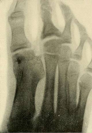
Pig. 122.—Malunion of Fracture of Large Toe, Three Years after theInjury, Causing Considerable Pressure. phalanges appear from the second to the ninth year, and unite withthe diaphysis between the sixteenth and the twenty-second year. Injuries and diseases of the phalanges are, of course, easilyrecognised. For a general view the tubal focus should be directlyabove the first phalanx of the middle toe. For differentiationfrom arthritis and chronic inflammatory processes skiagraphy ismost important. The time of exposure should not exceed half aminute—even in a few seconds useful reproductions of the toescan be obtained under favourable circumstances. CHAPTEK XISHOULDER AND UPPER EXTREMITY Shoulder.—The shoulder is fluoroscoped best while the patientis seated on a chair. (Compare section on the Position of thePatient, page 59.) Skiagraphy may be clone in the sitting, re-cumbent, and abdominal position. A table is less convenient forskiagraphing of the shoulder than the carpeted floo
Tags
Datum
Quelle
Copyright-info


