Similar
American quarterly of roentgenology (1909) (14570737930)
Zusammenfassung
Identifier: americanquarterl02amer (find matches)
Title: American quarterly of roentgenology
Year: 1909 (1900s)
Authors: American Roentgen Ray Society
Subjects: Nuclear Medicine Radiography Radiology Radiology
Publisher: Pittsburgh : American Roentgen Ray Society
Contributing Library: Francis A. Countway Library of Medicine
Digitizing Sponsor: Open Knowledge Commons and the National Endowment for the Humanities
Text Appearing Before Image:
e past decade and upon which discussionand criticism is invited from the members for mutual profit. Rachitis, Barlows disease, Acromegaly, Cretinism andMyxedema will not be discussed in this article as the usualmethods of diagnosis are quite satisfactory in above lesionsand their additional presentation would extend the scope ofthis paper so as to make it too lengthy. The lesions which will be shown in sequence are asfollows: Periostitis, enchondroma, ostitis, osteo-myelitis,osteo-malacia, ossium fragilitas, bone-cysts, osteoma, includingLone changes in early arthritis deformans, osteo-sarcoma andlastly tubercular, syphilitic and gonorrhoeal bone lesions. The first slide represents a common periosteal lesion foundafter typhoid fever—bulging out of the periosteal shadow andpractically no involvement of the bone itself. Simple peri-ostitis is difficult to diagnose until the inflammatory process *Read before the American Roent*en-Rav Society, Atlantic City, N. J.,September :23rd, 1909.
Text Appearing After Image:
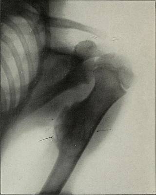
Fig. 9—Osteoma, verified microscopically. Diefenbach : X-Ray DIAGNOSIS L69 has produced some swelling and deposits of some size arenoted. This requires at least one week when circumscribedareas of darkness co-incident with exudation and sclerosisappear. Suppuration and necrosis are readily distinguishedwhen supervening- upon simple periostitis. In these casesthere are circumscribed areas of darkness and mottling- whichmay eventually extend over a large area. Dental injuries frequently produce maxillary periostitisand the Roentgen-ray will readily differentiate this lesionfrom neuralgia and neuritis at this and other locations. Anumber of cases of so-called facial or maxillary neuralgia were,after Roentgen examination, found to be due to periostealinflammation or abnormalities of the tegumentary system andnot due to nerve lesions. In periostitis gummosa infiltration of the gummatousdeposits causes a swelling- of the periosteum and of theHaversian canals; the nutrient marrow channels
Tags
Datum
Quelle
Copyright-info


