Similar
American quarterly of roentgenology (1906) (14570595450)
Zusammenfassung
Identifier: americanquarterl01amer (find matches)
Title: American quarterly of roentgenology
Year: 1906 (1900s)
Authors: American Roentgen Ray Society
Subjects: Nuclear Medicine Radiography Radiology Radiology
Publisher: Pittsburgh : American Roentgen Ray Society
Contributing Library: Francis A. Countway Library of Medicine
Digitizing Sponsor: Open Knowledge Commons and the National Endowment for the Humanities
Text Appearing Before Image:
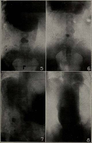
ce ofa non-palpable cancer which involved the stomach, thebismuth mixture persistently refusing to pass beyondthe blase and the tube meeting with a decided obstructionafter it had entered the stomach, the diagnosis of hour-glass malignant contraction would have seemed reason-able had not the further examination by means of thefull bismuth meal as illustrated by these negatives dis-proved this theory and explained the misleading data,while at the same time it pointed to the pylorus as thecause of the enormous dilation and the seat of thegrowth. Gastro-enterostomy, followed next day by apost-mortem examination, proved the entire correctnessof this view, and emphasized the importance of notmistaking the bottom of the blase for an organic stric-ture. Patient a young man, on right side. Dorso-ventralexposure. Gas occupies upper (left) part, bismuth thelower (right or pyloric) end. Notice spleen and leftkidney, whose upper border was palpable. Right dia-phragm considerably higher than left.
Text Appearing After Image:
To Illustrate the Presidents Address OF ROENTGENOLOGY. 9 Same patient on his belly. Presence of some foodprevents the bismuth from reaching quite to the bottomof the stomach. Lowest point of lesser curvature oppo-site second lumbar vertebra. The orthodiagraph showedthe lowest point of greater curvature one handbreadthbelow the navel. (No. 3.) Same patient on his back. Ventro-dorsal exposure.The fundus being lowered in this position, gravity servesto fill it out with bismuth. A girl, chlorotic, aet. 13, on her belly. Dorso-ventralexposure. Notice the sphincter of the antrum, the an-trum itself filled with bismuth, with the pylorus and apart of the duodenum below. Caudal pole above thenavel. Orthodiagraphy, the patient standing, locatedcaudal pole three finger-breadths below the navel. (No. 4.) Same patient on her back. Dorso-ventral exposure.Fundus better filled with bismuth than with patient onher belly. Part of duodenum clearly shown. Noticethat the caudal pole is considerably higher
Tags
Datum
Quelle
Copyright-info


