Similar
A system of instruction in X-ray methods and medical uses of light, hot-air, vibration and high-frequency currents - a pictorial system of teaching by clinical instruction plates with explanatory text (14756662312)
Zusammenfassung
Identifier: systemofinstruct00mone (find matches)
Title: A system of instruction in X-ray methods and medical uses of light, hot-air, vibration and high-frequency currents : a pictorial system of teaching by clinical instruction plates with explanatory text : a series of photographic clinics in standard uses of scientific therapeutic apparatus for surgical and medical practitioners : prepared especially for the post-graduate home study of surgeons, general physicians, dentists, dermatologists and specialists in the treatment of chronic diseases, and sanitarium practice
Year: 1902 (1900s)
Authors: Monell, S. H. (Samuel Howard), d. 1918
Subjects: Vibration X-rays Diagnosis, Radioscopic Thermotherapy Electrotherapeutics X-Ray Therapy Vibration Diagnosis
Publisher: New York : E.R. Pelton
Contributing Library: Francis A. Countway Library of Medicine
Digitizing Sponsor: Open Knowledge Commons and Harvard Medical School
Text Appearing Before Image:
the depth ofthe bullet in the tissues. The use of this device is illustrated in In-struction Plates No. 78 to 83 inclusive. The authors description isas follows: * Suppose a rectangular plane. Assume that the upper edge ofthe rectangle intersects two foci producing X-rays. The rays fromone focus cross those proceeding from the other; but it is always pos-sible to determine to which tube any one of the rays belongs, whatevermay be the point in the plane at which we place ourself. Let us now neglect all that part of the plane touching the tubes;the direction of the rays will then be represented only by lines of afew centimetres in length, but by prolonging these lines we will reachthe centre of each of the foci. Let us now interpose between the tubesand the remaining part of tlie j)lane a fluoroscent screen, and interposean opaque body to the X-rays, the shadow of which is thrown on theluminous screen. If the foreign body is in this plane, its shadow will * Archives of the Roentgen Ray.
Text Appearing After Image:
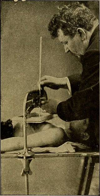
Plate 78.—The Remy Localizer. First ijo.sitioii. With tlie tirst tube below throwingthe shadow of the object on the screen seen on the chest of this patient push down the firstrod in the axis of the rays till its point marks the centre of the shadow. This rod thenresembles one of the threads in the more familiar cross-thread localizer.
Tags
Datum
Quelle
Copyright-info


