Similar
A system of instruction in X-ray methods and medical uses of light, hot-air, vibration and high-frequency currents - a pictorial system of teaching by clinical instruction plates with explanatory text (14754694324)
Zusammenfassung
Identifier: systemofinstruct00mone (find matches)
Title: A system of instruction in X-ray methods and medical uses of light, hot-air, vibration and high-frequency currents : a pictorial system of teaching by clinical instruction plates with explanatory text : a series of photographic clinics in standard uses of scientific therapeutic apparatus for surgical and medical practitioners : prepared especially for the post-graduate home study of surgeons, general physicians, dentists, dermatologists and specialists in the treatment of chronic diseases, and sanitarium practice
Year: 1902 (1900s)
Authors: Monell, S. H. (Samuel Howard), d. 1918
Subjects: Vibration X-rays Diagnosis, Radioscopic Thermotherapy Electrotherapeutics X-Ray Therapy Vibration Diagnosis
Publisher: New York : E.R. Pelton
Contributing Library: Francis A. Countway Library of Medicine
Digitizing Sponsor: Open Knowledge Commons and Harvard Medical School
Text Appearing Before Image:
Plate 147.—Measuring Pulmonary Shadows by the Skiameter. Dorsal position. Holdthe Skiameter over successive areas of the chest while observing the shadows from behind witha large mounted open screen instead of the fluoroscope. Cut does not show the light-screen tohide the tube or the use of the cloth which may be thrown over the head and around the fieldof examination to create a dark chamber for the eyes. Both plates teaching the use of theSkiameter were photographed especially for this work by Dr. A. W. Crane, of Kalamazoo.
Text Appearing After Image:
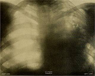
W 2 SD S 2 o X-RAY DIAGNOSIS IN DISEASES OF THE CHEST 345 the most opaque tissue of the body. With this assertion I cannotagree. Look, for instance, at the picture now on the screen. Itrepresents a portion of a rib, a piece of muscle the same size, and ablood-clot. My eye can detect no difference between the muscle andthe blood-clot—that is to say, clotted blood, which, by the bye, is moreopaque than fluid blood, is no more opaque to the rays than muscle.Again I took the lung and kidney which contained tubercle fromthe same case, and radiographed them together. The tubercle in thekidneys is distinctly seen, down to the minutest miliary tuberclewhich could be detected by the naked eye. Again, look at theselungs from a case of acute miliary tuberculosis. They show thetubercle scattered through their substance clearly enough. Puttingthe above facts together, we may, I think, answer our first questionby saying that the Roentgen rays. can show definitely tubercle inthe lung. I will now t
Tags
Datum
Quelle
Copyright-info


