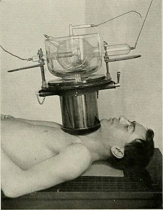
Röntgen rays and electro-therapeutics - with chapters on radium and phototherapy (1910) (14571511490)
Summary
Identifier: rntgenrayselectr00kass (find matches)
Title: Röntgen rays and electro-therapeutics : with chapters on radium and phototherapy
Year: 1910 (1910s)
Authors: Kassabian, Mihran Krikor, 1870-1910
Subjects: Electrotherapeutics X-rays Phototherapy Radiology Radiotherapy
Publisher: Philadelphia & London : J.B. Lippincott Company
Contributing Library: Francis A. Countway Library of Medicine
Digitizing Sponsor: Open Knowledge Commons and Harvard Medical School
Text Appearing Before Image:
'
Text Appearing After Image:
THE CLINICAL APPLICATIONS. 265 are of great diagnostic aid, as the contour of these bones is very irregularand the rays must traverse great density of structure. Fractures of thepelvic bones are divided into those in which the individual parts arefractured and those in which the pelvic rim is broken. In skiagraphing the pelvis the patient must assume the ventral anddorsal decubitus positions. In a skiagraph of the sacro-coccygeal regionthe tube should be placed over the umbilicus so that the shadow of thepubic symphysis will not overlap the shadow of the sacrum or coccyx.The rectum should be emptied by an enema prior to the examination. The ilium, ischium, and the pubes can be skiagraphed in the abovemanner, with slight modifications in the relation of the tube, the part,and the plate. The Spinal Column. For the sake of conveniently studying the spinal column, it is dividedinto the cervical, dorsal, and lumbar regions. The cervical region is best skiagraphed in lateral view. Completef
Tags
Date
Source
Copyright info


