Similar
Röntgen rays and electro-therapeutics - with chapters on radium and phototherapy (1910) (14755899544)
Summary
Identifier: rntgenrayselectr00kass (find matches)
Title: Röntgen rays and electro-therapeutics : with chapters on radium and phototherapy
Year: 1910 (1910s)
Authors: Kassabian, Mihran Krikor, 1870-1910
Subjects: Electrotherapeutics X-rays Phototherapy Radiology Radiotherapy
Publisher: Philadelphia & London : J.B. Lippincott Company
Contributing Library: Francis A. Countway Library of Medicine
Digitizing Sponsor: Open Knowledge Commons and Harvard Medical School
Text Appearing Before Image:
used. Theshadows thrown by the last ribsand the transverse process of thefirst lumbar vertebrae are to betaken as guides. If nothing is seen at the first attempt, it should not be concluded that the result isnegative. The plate should be intensified, and allowed to dry. Thisbrings out many shadow details, previously invisible. To obtain the besteffects the plate should be examined at a distance of 5 or 6 ft. If anyspecks are seen, which may possibly be due to calculi, another exposureshould be made within three or four days. In any case of doubt a sepa-rate exposure should be made. A lead pipe with an opening of 13 cm. indiameter is placed close to the tube, and 50 cm. from the plate, so asto cut off the secondary rays and obtain a well-defined shadow. (Figs.192 and 193.) My time of exposure in renal skiagraphy depends uponthe corpulence of the patient, and the degree of high penetrative powerof the tube. The distance of the target from the plate is from 22 to 30inches (55 to 75 cm.).
Text Appearing After Image:
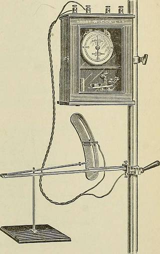
Fig. 191.—Clock arrangement and break of the same. 356 ELECTEO-THEEAPEUTICS. The metliod of examination of the kidney by the X-rays, when theorgan is outside of the body during operation, has been fully describedby the discoverer, Mr. Fenwick.^ It consists in examining the kidneywith the fluorescent screen after the organ has been removed as far aspossible from the abdominal cavity. In some cases, he says, the kidneycannot be displaced out far enough to permit of a screen examination,due to insufficient length of the renal vessels. An objection to thismethod of examination is that the surgeon must necessarily remainin darkness for at least ten or fifteen minutes before he will be able tosuccessfully perform a screen examination. F. Voelcker and A. Lichtenberg ^ describe a process of pyelography.The ureter is catheterized, and the instrument is advanced to the renalpelvis. A 5 per cent, solution of a silver salt is then slowly injectedthrough the catheter. There are individual variat
Tags
Date
Source
Copyright info


