Similar
Röntgen rays and electro-therapeutics - with chapters on radium and phototherapy (1910) (14757887092)
Summary
Identifier: rntgenrayselectr00kass (find matches)
Title: Röntgen rays and electro-therapeutics : with chapters on radium and phototherapy
Year: 1910 (1910s)
Authors: Kassabian, Mihran Krikor, 1870-1910
Subjects: Electrotherapeutics X-rays Phototherapy Radiology Radiotherapy
Publisher: Philadelphia & London : J.B. Lippincott Company
Contributing Library: Francis A. Countway Library of Medicine
Digitizing Sponsor: Open Knowledge Commons and Harvard Medical School
Text Appearing Before Image:
tages of Pottsdisease from intercostal neuralgia, renal disease, empyema with subdia-phragmatic abscess, etc.; but the skiagram will show the bodies of thevertebrae and the interarticular spaces to possess a denser shadow thannormal. In advanced cases the disintegrated osseous tissue will present adark, dense, irregular shadow. Place the patient in the dorsal decubitusposition, have him flex the knees so as to straighten the spine as far aspossible and thus bring it in closer relation with the plate. The abovedescription applies to any region of the spine. Dark shadows in theright iliac fossa, often due to the accumulation of gases in the colon,must not be mistaken for necrosis of bone. Amputation Stumps.—The process of healing can be systematicallyfollowed in cases of amputation stumps, by noting the existence or ab-sence of a fine layer of compact bony tissue, covering the medullary canal,and thus the presence of a sequestrum, interfering with the healing, canlikewise be detected.
Text Appearing After Image:
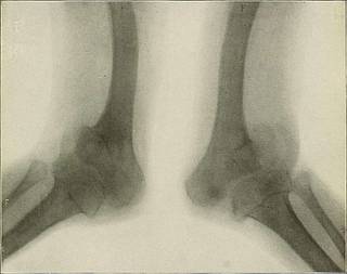
Fig. 159.—Delayed Ossification of the Epiphyses.—Patient 55 years of age. Every bone deformed.Unable to \yalk since childhood and had been in the hospital more than 30 years. No history of syphilis, andDr. Burr of the Philadelphia Hospital believes the deformities to be congenital and due to disease of the spinalcord which developed during fcetal life. The epiphyseal ends of the femora, tibise, and fibulae look spongy fromlack of ossification. Articular surfaces irregular, bones bent and pervious to the rays. The epiphyseal linesappeared darker because of excessive ossification. THE CLINICAL APPLICATIONS. 271 Resection of Joints.—Before resecting a joint, the rays will determinetlie exact character of the affection, and their application after the woundhas been dressed will inform the operator if the bones are in the bestpossible position. Regeneration of Bone.—After removal of a portion of bone, the peri-osteum being left intact, the formation of new bone may be carefullyobse
Tags
Date
Source
Copyright info


