Similar
Radiography, X-ray therapeutics and radium therapy (1916) (14758280395)
Summary
Identifier: radiographyxrayt00knox (find matches)
Title: Radiography, X-ray therapeutics and radium therapy
Year: 1916 (1910s)
Authors: Knox, Robert, 1868-1928
Subjects: Radiography Radiotherapy Radium
Publisher: New York : Macmillan
Contributing Library: University of California Libraries
Digitizing Sponsor: Internet Archive
Text Appearing Before Image:
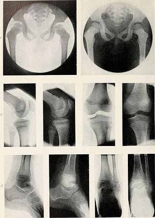
Fig. 115.—Lateral view of knee-joint, showing epiphyses.Note prolongation of tibial epiphysis on anterior aspectof tibia. Age, 11 years. The epiphyseal line is seen atthe level of theadductor tubercleon the inner side.It is wavy in out-line, rises sharplytowards the centre,and has a slightlylower level at theouter side of thebone. The epiphysesof the tibia andEpiphyseal fibula will be seenin the picture. Theepiphyseal line ofthe tibia resemblesthat of the line ofthe lower end ofthe femur. Theupper epiphysis ofthe fibula is a smallmass, appearing torest on the top ofthe shaft.
Text Appearing After Image:
PLATE IX.—Showing Epiphyses of Hip, Knee, and Ankle Joints. «, Pelvis and hip-joints in a child of 5-6 years. Knee-joint in a child 10-12 years, b, Lateral view, c, Anteroposterior view. Ankle-joint in a child 10-12 years. /, Lateral aspect, e, Antero-posterior aspect. THE KNEE-JOINT 147 In a lateral view the epiphyseal lines of the femur and fibula are nearly
Tags
Date
Source
Copyright info


