Radiography, X-ray therapeutics and radium therapy (1916) (14735366526)
Zusammenfassung
Identifier: radiographyxrayt00knox (find matches)
Title: Radiography, X-ray therapeutics and radium therapy
Year: 1916 (1910s)
Authors: Knox, Robert, 1868-1928
Subjects: Radiography Radiotherapy Radium
Publisher: New York : Macmillan
Contributing Library: University of California Libraries
Digitizing Sponsor: Internet Archive
Text Appearing Before Image:
rt tubes called calices, which surround the renal papillaeat the bottom of the sinus. These join each other, with or without theintervention of short passages called infundibula, to form usually two tubes,the upper and lower pelves, and the union of the two pelves constitutes thecommon pelvis renales, which generally narrows to the size of a goose quill,and becomes the ureter proper. The ureters pierce the bladder at thejunction of the posterior or lateral walls, about an inch and a half above thebase of the prostate. The left ureter is contained in the root of the posteriorfalse ligament of the bladder (or in part of the broad ligament in thefemale), and can be traced beneath the peritoneum to its entrance into thefundus of the bladder. Diseases of Urinary Tract In order to make a diagnosis from negatives of the urinary tract indisease, it is necessary for the radiographer to be familiar with the appearanceof good normal negatives from the region of the kidneys, ureters, and bladder.
Text Appearing After Image:
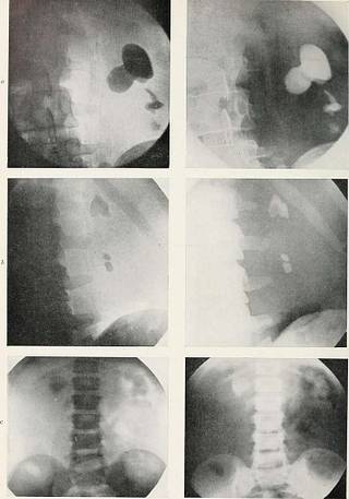
PLATE XLix.—Uhinahy Calculi. a, Calculi in kidney. b, Calculi in kidney. (Radiograph by C. Thurston Holland.) c, Faecal mass in kidney area simulating calculus. CALCULUS IN THE KIDNEY 241 This knowledge can only be acquired by regular practice, though it is possibleto demonstrate the essential points by means of a series of radiographs. Agood radiograph of the kidney area should show the outline of the organ,and should cover the whole of the kidney. In order to get the whole of thearea, it is necessary to get the two lower ribs in the picture. Bearing in mind the normal appearances of the urinary tract, we nowproceed to a consideration of the abnormalities which may be met with inthe investigation of diseases of the urinary organs. Before considering those .diseases in order, it is necessary to consider some of the conditions which,when met with, are apt to mislead the observer and cause errors of diagnosis.Those are numerous and ever increasing in number as fresh cases are recorded
Tags
Datum
Quelle
Copyright-info


