Similar
Radiography, X-ray therapeutics and radium therapy (1916) (14571655240)
Zusammenfassung
Identifier: radiographyxrayt00knox (find matches)
Title: Radiography, X-ray therapeutics and radium therapy
Year: 1916 (1910s)
Authors: Knox, Robert, 1868-1928
Subjects: Radiography Radiotherapy Radium
Publisher: New York : Macmillan
Contributing Library: University of California Libraries
Digitizing Sponsor: Internet Archive
Text Appearing Before Image:
orts for removal may be facilitated by a screenexamination, and the forceps guided to the tube. Irregularities in the Outline of the Diaphragm, to whatevercause they may be due, will often give appearances which lead to difficultiesin diagnosis ; moreover, a secondary involvement of the liver is not at alluncommon in cases where a growth in the lung exists. Tumours of the chest ivall may also have to be excluded. Hydatid cyst, though not common in this country, must not be over-looked. The appearance of such a cyst is diagnostic, and a rounded, sharply-cut shadow in any part of the lungs should excite suspicion of hydatiddisease. Cysts may arise from any of the structures composing the walls,i.e. sternum, ribs, costal cartilages, or spine. The appearances presentedby a case of primary sarcoma arising from the inner aspect of a rib are typicalof cyst—a rapidly growing sarcoma, which becomes hemorrhagic, the wallsof the growth consist of thickened pleura, and the cavity is filled with
Text Appearing After Image:
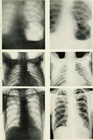
PLATE XXXIX.—Chests. a, The arch of the diaphragm on the left Bide is high, the clear area is caused by gas in a distended■stomach. Note fluid level at the lower limit of the clear area. b, Extensive distribution of calcareous glands in thorax, axillae, and cervical regions. Eealed tubercu-losis of many years standing. c, Calcilieil ^laml* at roots otliotli lungs, lh-aletl tuberculosis. EXAMINATION OF THE HEART AND AORTA 205 blood-clot and growth. The X-ray appearances will show it to have a well-defined wall with semi-fluid contents. The Examination of the Heart and Aorta Variations in the Size, Shape, and Position of the Heart.—Theheart may be greatly enlarged in all directions, or it may show a markedhypertropny of the left ventricle. Dilation of the right side of the heartmay be distinguished from hypertrophy by the lack of density in the shadows. The heart may be displaced to one or other side. It is sometimes seenon the right side of the thorax, the aorta in such a case be
Tags
Datum
Quelle
Copyright-info


